Facilities
Optical and Confocal Microscopy

The Microscopy Service offers microscopes capable of working with diverse types of illumination, detection and acquisition that allow working with different applications and samples.
- Brightfield transmitted light microscopy
- Conventional fluorescence microscopy
- Advanced brightfield and fluorescence microscopy
- Laser scanning confocal microscopy
- Laser-based Microdissection with brightfield and fluorescence
- Total Internal Reflection Fluorescence (TIRF) microscopy and Super-resolution (STORM).
- Brightfield transmitted light microscopy
- Brightfield Microscopy
- Phase contrast Microscopy
- Nomarski differential interference contrast microscopy (DIC).
Brightfield microscopy is based on the transillumination of simples using a white light source, focused through the condenser in order to pass through the sample into the objective lens that collects light for visualization through the eyepieces or acquisition using a digital camera. This technique is useful for visualizing cells, tissues, colorimetric histological sections, animal models (zebra fish, C. elegans). Phase contrast and Nomarski are brightfield visualization methods using lenses and specific optical elements with improve the contrast of structures with would otherwise be invisible.
- Conventional fluorescence microscopy
- Upright microscopes with mercury lamps (or similar)
- Inverted microscopes with mercury lamps (or similar)
Fluorescence is the process of the absorption de un photon y subsequent emission of another photon with less energy (longer wavelength) by a molecule (Fluorochrome). Conventional fluorescence combines a powerful white light source with optical filters and mirrors to achieve specific excitation and detection wavelengths for each fluorophore. Using labelling techniques, we can combine different fluorophores to mark different cell types and subcellular regions (plasma membrane, nucleus etc.).
The IBiS offers various conventional microscopes that are apt for the visualization of fluorescent histological sections, fixed cultured cells and animal models. Most microscopes can work with blue (DAPI, Hoechst), green (GFP, FITC, Alexa fluor 488), and red (TRITC, Alexa fluor 555) fluorophores. For compatibility with other markers, refer to the Equipment page for detailed specifications or consult the support staff.
- Advanced brightfield and fluoroesence microscopy
- Thunder 8 channel LED high speed microscope
- newCAST Stereology microscopes
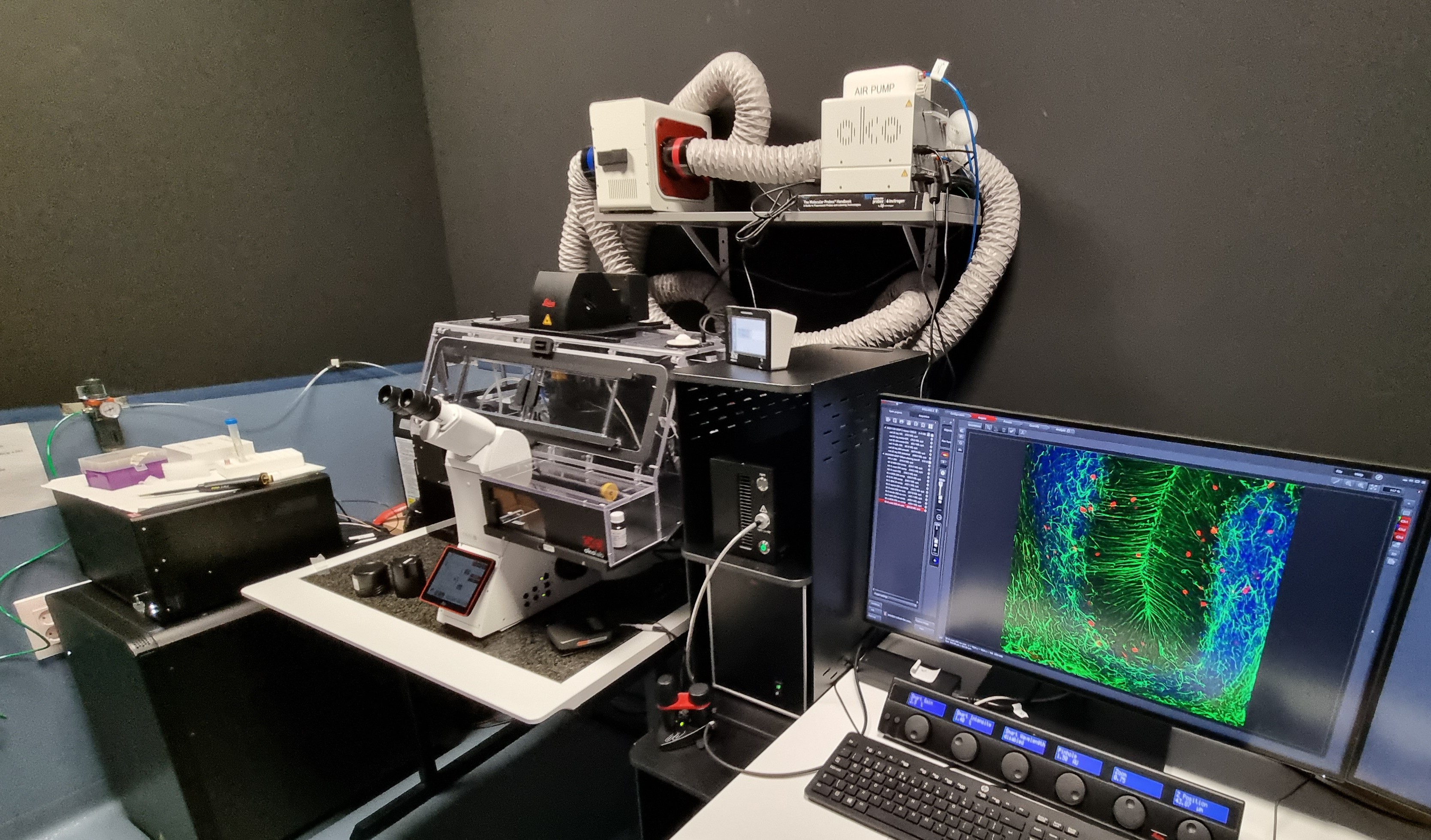
In addition to its conventional microscopes, the IBiS possesses advanced microscopes that combine software and hardware improvements to be able to acquire better quality images, with high speed and with more quantitative information.
The Leica Thunder microscope combines hardware optimized for very high-speed acquisition with automatic image processing. The result is a system that can work as an effective slide scanner with histological samples or cell culture. It is compatible with brightfield, phase contrast, colorimetric labeling and up to 8 high quality fluorescence channels.
NewCAST microscopes offer the automatic acquisition of large image mosaics of histological samples, both brightfield and fluorescence, with digital stereology tools that make it possible to obtain unbiased data (e.g., area or volume) sampling small areas of the sample and extrapolating the results in a statistically robust manner,
- Laser scanning confocal microscopy
- Microscopio Confocal Leica SP2
- Microscopio Confocal Nikon A1
- Microscopio Confocal Leica Stellaris 8
Confocal microscopy uses optical methods to improve contrast, remove unfocused light and obtain images as optical slices making it possible to generate 3D reconstructions. These systems are typically used with diverse types of fluorescently labelled samples but it also possible to use reflected light for unlabeled samples in applications such as dentistry, materials and even live tissues (e.g., cilia).
- Laser-based Microdissection with brightfield and fluorescence
Laser microdissection is a method of isolating specific regions of tissues or cell culture for subsequent analysis using genomic or proteomic techniques. Samples are mounted between glass slide and a membrane that enables their isolation without contamination from external environmental impurities. Once the region is selection, the sample is cut automatically via software using a high precision laser beam. Single cell or tissue region isolation is valuable for many research fields such as Molecular Pathology, Cellular Biology, Cancer research, Forensic medicine, Neuroscience, and Food science.
- Total Internal Reflection Fluorescence (TIRF) microscopy and Super-resolution (STORM)
Total Internal Reflection Fluorescence (TIRF) microscopy takes advantage of a physical phenomenon which allows excitation laser light to be angled such that with reflects completely within the coverslip/glass-bottom and only illuminates the sample in the region very close (~200 nm) to the coverslip. This method is very useful for studies into the plasma membrane, receptor dynamics, integrins and the cytoskeleton. It also permits single molecule tracking, giving high contrast and high axial resolution with very low phototoxicity and photobleaching. TIRF is often combined with Super-resolution (STORM) which exploits the stochastic activation of individual fluorophores to obtain a lateral resolution of up to 20 nm.
Key applications and services
- Colocalization of fluorescent markers as evidence for function interaction.
- FRET (Förster resonance energy transfer) allows direct protein-protein interactions to be detected by detecting energy transfer between fluorophore in the angstrom range.
- FRAP (fluorescence recovery after photobleaching) experiments allow molecular dynamics to be studied (movable vs immovable fractions)
- Fluorescence lifetime microscopy (FLIM) allows fluorochromes to be distinguished by their lifetime properties (in addition to excitation and emission). FLIM can also be used for functional studies such as FLIM-FRET, which can give more robust measurements than simple intensity-based approaches.
- 3D reconstruction and quantification.
- In vivo experiments using live samples with microscope enclosures with temperature, humidity, O2 and CO2 control.
- Experimental design advice – contact us for assistance in sample processing, staining and choice of microscopy technique.
Donate
Promote science with a donation and be part of the change.


